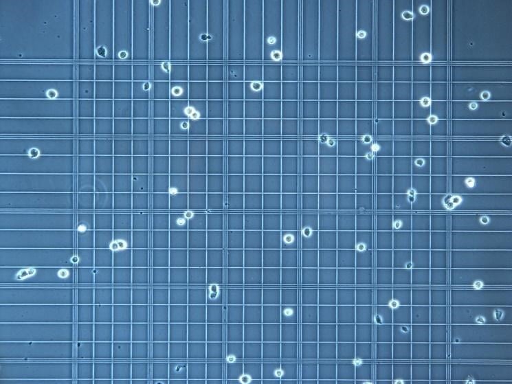
Dye exclusion assays, such as the Trypan Blue assay, are rapid and straightforward methods used to qualitatively (and often semi-quantitatively) assess cell viability based on membrane integrity. The principle relies on the inability of viable cells with intact cell membranes to take up certain dyes, while non-viable cells with damaged membranes allow the dye to enter and stain the cytoplasm. This results in a visual distinction between unstained (live) and stained (dead) cells, typically observed under a light microscope.
Test Details
Procedure
- Cells were harvested following treatment or experimental manipulation, and a suspension of the cells was prepared.
- A dye solution (e.g., Trypan Blue) was mixed with the cell suspension.
- A small aliquot of the mixture was then placed on a microscope slide and examined under a light microscope to count the number of stained (non-viable) and unstained (viable) cells.
- Freshney, R. I. (2010). Culture of animal cells: a manual of basic technique (6th ed.). John Wiley & Sons. (Provides a general overview of cell viability assays, including dye exclusion).
- Strober, W. (2001). Trypan blue exclusion test of cell viability. Current Protocols in Immunology, Appendix 3, A.3B.1-A.3B.3. (A detailed protocol for the Trypan Blue assay).
Related Devices
-
CO2 Cell Culture Incubator
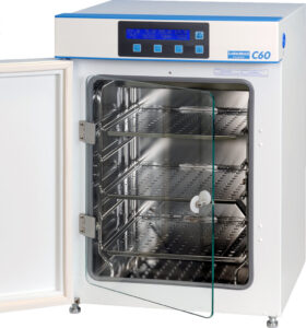 A carbon dioxide (CO2) incubator is a specialized laboratory equipment used for cell culture, which helps to maintain optimal & precise conditions of temperature, humidity, and carbon dioxide levels required…
Contact Us
Have questions about this test? Send us a message and we'll get back to you as soon as possible.