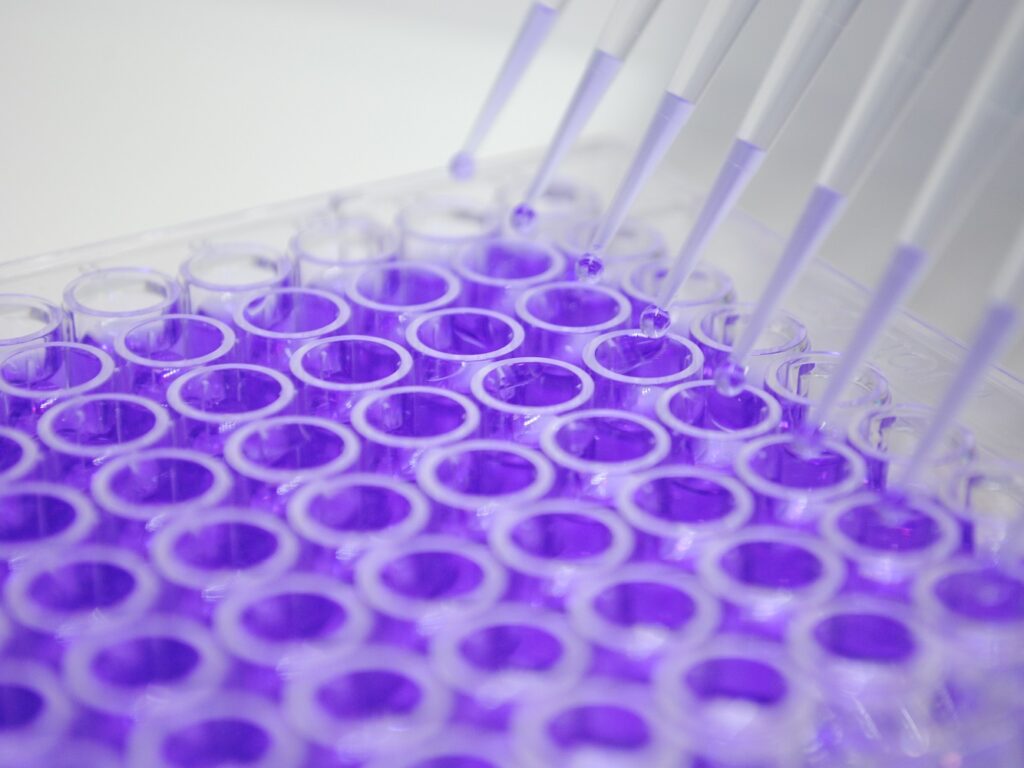
The crystal violet assay is a straightforward and widely used colorimetric technique for the quantitative determination of adherent cell number or biomass. It relies on the ability of crystal violet, a cationic dye, to bind electrostatically to negatively charged components of cells, including DNA and proteins. Following staining and washing to remove unbound dye, the dye bound to the cells is eluted using a solubilizing solution. The optical density of the resulting solution is then measured spectrophotometrically, providing a quantitative measure of the number of cells present in the culture. This assay is valuable for assessing cell proliferation, cytotoxicity, and cell adhesion in various biological studies.
Test Details
- The experiment began with cell cultivation. Cells were seeded in vessels, allowed to attach and grow. They were then treated according to the design and washed to remove debris.
- Next, the treated cells were prepared for analysis. Fixation preserved their structure, followed by staining with crystal violet to bind cellular components. Unbound stain was thoroughly washed away.
- Finally, the bound stain was quantified. Crystal violet was extracted from the cells using a solution, and its absorbance was measured. This reading determined the cell number or biomass at the experiment's conclusion
- Kueng, W., Silber, E., & Eppenberger, U. (1989). Quantification of cells cultured on 96-well plates. Analytical Biochemistry, 182(1), 16-19.
- Freshney, R. I. (2010). Culture of animal cells: a manual of basic technique (6th ed.). John Wiley & Sons.
Related Devices
-
CO2 Cell Culture Incubator
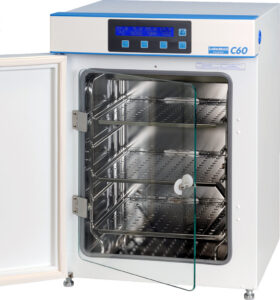 A carbon dioxide (CO2) incubator is a specialized laboratory equipment used for cell culture, which helps to maintain optimal & precise conditions of temperature, humidity, and carbon dioxide levels required…
 -
ELISA Plate Reader
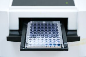 The ELISA reader is a specialized spectrophotometer—an apparatus for measuring the intensity of light throughout a part of the spectrum—that measures the reactions of antigens and antibodies in a solution…
Contact Us
Have questions about this test? Send us a message and we'll get back to you as soon as possible.